Medically reviewed and approved by a board-certified member
Zoology
COMPARATIVE ANATOMY: DIGESTIVE SYSTEM OF BIRD, MAMMAL AND REPTILE
By BS MediaTwitter Profile | Updated: Sunday, 09 July 2017 13:44 UTC
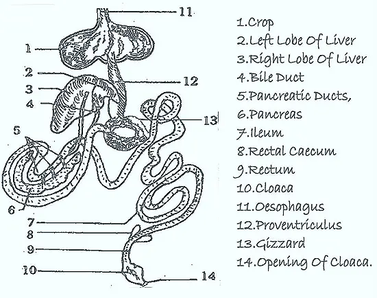
Digestive System Of Bird: Eg: Pegion Digestive System (Columba)
Digestive System Of Mammal: Eg: Rabbit Digestive System (Oryctolagus)
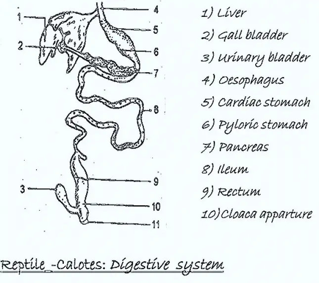
Digestive System Of Reptile: Eg: Lizard Digestive System (Calotes)
In previous topic we discussed about the comparative anatomy of digestive system of frog and shark. In this topic we discussed about the comparative anatomy of digestive system of Bird, Mammal and Reptiles.
Calotes is a poikilothermic and terrestrial lizard. Columba and Oryctolagus are warm blooded animals. Columba is adapted for mode of life-bird. Oryctolagus (Rabbit) is a mammal. The digestive system consists of alimentary canal and its associated digestive glands. The living of the alimentary canal is mostly endodermal in origin being derived from the wall of the archenteron. Distinc' salivary glands secreting enzymes are present only in mammals. In reptiles, the oral glands are present in various positions termed labial, parotid, lingual & sublingual etc. The secretions of which serve primarily to keep the mouth moist and secondarily to facilitate the movements of the tongue. The other connected with the midgut and arising as out growth are the liver and pancreas.
DIGESTIVE SYSTEM OF BIRD
| Calotes (Lizard) | Columba (Pigeon) | Oryctoiagus (Rabbit) |
| 1. Mouth is a wide, slit present at the anterior end of head. | 1. Mouth is terminal, slitlike aperture bounded by horny Jaws. | 1. Mouth is sub-terminal, cleft bounded bv mobile, fleshy lips. |
| 2. Buccal cavity is a narrow gap. Labial glands are present on lips.They secrete mucous. | 2. Buccal cavity is narrow and some what and dorsoventrally flattened. | 2. Buccal cavity is a spacious chamber andits space between lips and the teeth is called vestibule. This receives the mouth opening. |
| 3. On both jaws teet are present, polyphyodont homodont teeth arranged in a single row on each jaw. Teeth are not useful for mastication. Pleurodont dentition is present. | 3. Jaws are modified into tooth less beak. | 3. Dentition in mammals:Teeth are diphyodont, heterodont and thecodont. These are arranged in a single row on each Jaw.Teeth are modified cutting (Incissors) and chewing (Premolars & molars) canines are absent in rabbit. |
| 4. Tongue is attached posteriorly to the floor of buccal cavity and is free anteriorly. Sensory papillae are present. | 4. Tongue is narrow triangular and fleshy. Its surface is covered with horny material and bears thorn-like projections which carry taste buds and mucous glands. | 4. Tongue is highly specialized, fleshy and muscular and can be moved in different directions. It can be protruded out. Its surface is rugose being covered with numerous papillae along with taste buds. |
| 5. A pair of internal nostrils open into the roof of the buccal cavity anteriorly. Hard palate is present. | 5. A bony palate is wanting in birds but a pair of palatal folds and palatal groove between the two folds are present. Internal nostrils are located dorsal to the palatal folds. | 5. The nasal passages are separated from the buccal cavity by a bony palate. The internal nostrils open into the pharynx nearer to glottis. |
| 6. A bony palate is present covering the roof of the buccal cavity. | 6. A bony palate is absent. But soft palate is formed of two membranous folds. | 6. The palate is differentiated into anterior bony hard palate and a soft palate is formed of connective tissue. The soft palate is produced behind into a process – velum palati hanging down from the roof, which prevents the entry of food into nasal passage. |
| 7. Unicellular mucous glands are present and keep the buccal cavity always wet. | 7. Unicellular mucous glands are absent in the epithelium of bucco-pharyngeal region. | 7. Unicellular mucous glands are absent. But multi cellular serous glands are present. |
| 8. Salivary glands are absent. But labial glands are open at the lips which do not play any role in digestion. | 8. Salivary glands which open into the buccal cavity are lingual, mandibular, maxillary, cricoary tenoid, palatinal and sphenopalatinal glands. | 8. The multi cellular salaivary glands are four pairs. They are Infra orbital, parotid, sublingual and sub-maxillary glands. Palatine, tonsillar, superior & inferior labial glands are also associated. |
| 9. Pharynx is marked off. On the roof of pharynx near the junction of two jaws a pair of openings is called Eustachian apertures. The floor of pharynx has the glottis. | 9. Pharynx is marked off from the buccal cavity but it receives, internal nostrils through which nasal passages open into its cavity, the gullet & glottis. | 9. Pharynx is not sharply demarcated from the buccal cavity. It receives the openings of esophagus and the glottis. |
| 10. Esophagus is a narrow tube and straight extends through the neck. Mucous glands are present. | 10. Oesophagus is a bng and narrow tube. It has thick walls. Mucous glands are absent. | 10. Oesophagus is a long thin walled tube. It is clearly marked off from the pharynx as well as stomach. Mucous glands are present. |
| 11. Crop is absent. | 11. The oesophagus is dilated into a thin-walled sac the crop. It secrete pigeon milk in both sexes and used to feed the young birds. | 11. Crop is absent. The wall of oesophagus is produced into the cardiac stomach to form cardiac valve. |
| 12. Stomach is a sac- like structure. Its anterior part is cardiac stomach and posterior part is pyloric stomach. At the end of pyloric stomach a small constriction is present. It possesses a pyloric sphincter. | 12. Stomach is divided into a glandular proventriculus and posterior muscular gizzard. Gizzard acts like grinding apparatus. Pyloric valve absent. | 12. Stomach is divisible into cardiac, fundic and pyloric parts. Pyloric stomach contains pyloric valve. |
| 13. Intestine is differentiated into duodenum and ileum. Intestine very long and very much coiled because is a herbivorous animal | 13. Same structures are present. But the bile duct and pancreatic ducts open separately into the proximal and distal ends of the duodenum respectively. | |
| 14. A single rectal caecum is present. | 14. At the junction of ileum & colon, two divertulae are present. These are called Rectaicaeca. | 14. An ilio-colic valve is present at the junction of the small intestine and large intestine or colon. At the junction of these portions, a spiral shaped vermi form appendix is present. At the regular intervals of the colon shows pocket-like pouches - 'Haustra'.Colon shows longitudinal muscle folds taeniae'. Cellulose enzyme is produced. |
| 15. Cloaca is common opening for digestive'and urinogenital ducts. It is divided into coprodaeum, urodaeum, and proctodaeum. | 15. Cloaca is large and divided into the same parts. On the dorsal side of the proctodaeum a thick walled blindsa "Bursa fabricii" is present. It becomes degenerated in the adult and is known as cloacal thymus. | 15. Cloaca is absent. Anus is present. |
| 16. Liver is bilobed gland. The right lobe has a gall bladder. It secretes bile (alkaline& no enzymes). | 16. Liver is bilobed dark red gland. Gall Bladder is absent. Separate bile ducts are formed.Liver secretes bile. | 16. Liver is very large and consists of five lobes. Kupffer's cells are present in the liver. Liver secretes bile. Gall Bladder is present. |
| 17. Pancreas is a whitish gland present between stomach duodenum. | 17. Pancreas is a pink coloured gland present the loop of duodenum. Three pancre catic ducts open into the duodenum. | 17. pancreas is a diffused gland of pink colour. Pancreatic duct opens into the duodenum. |
| 18. Gastric, intestinal glands are also present. | 18. Same are present. | 18. Same are present. |
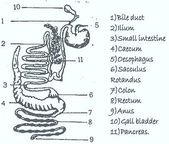
DIGESTIVE SYSTEM OF RABBIT
Tags:
End of the article