Medically reviewed and approved by a board-certified member
Hemotology
PRINCIPLES OF WORKING OF AUTOMATED HEMOTOLOGY ANALYZER
By Dayyal Dg.Twitter Profile | Published: Monday, 31 July 2017
Automated hematology analyzers work on different principles:
- Electrical impedance
- Light scatter
- Fluorescence
- Light absorption
- Electrical conductivity.
Most analyzers are based on a combination of different principles.
(1) Electrical impedance: This is the classic and timetested technology for counting cellular elements of blood. As this method of cell counting was first developed by Coulter Electronics, it is also called as Coulter principle (see Figure 811.1). Two electrodes placed in isotonic solutions are separated by a glass tube having a small aperture. A vacuum is applied and as a cell passes through the aperture, flow of current is impeded and a voltage pulse is generated.
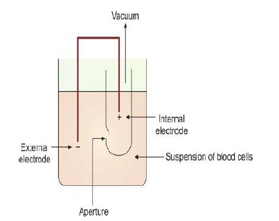
Figure 811.1 Coulter principle of electrical impedance
The requisite condition for cell counting by this method is high dilution of sample so that minimal numbers of cells pass through the aperture at one point of time. There are two electrodes on either side of the aperture; as the solution in which the cells are suspended is an electrolyte solution, an electric current is generated between the two electrodes. When a cell passes through this narrow aperture across which a current is flowing, change in electrical resistance (i.e. momentary interruption of electrical current between the two electrodes) occurs. A small pulse is generated due to a temporary increase in impedance. This pulse is amplified, measured, and counted. The height of the pulse is proportional to cell volume. The width of the pulse corresponds with the time required for the cell to traverse the aperture. Cells that do not pass through the center of the aperture generate a distorted pulse that is not representative of the cell volume. Some analyzers use hydrodynamic focusing to force the cells through the central path so that all cells take the same path for volume measurement.
An anticoagulated whole blood sample is aspirated into the system, divided into two portions, and mixed with a diluent. One dilution is passed to the red cell aperture bath (for red cell and platelet counting), and the other is delivered to the WBC aperture bath (where a reagent is added for lysis of red cells and release hemoglobin; this portion is used for leukocyte counting followed by estimation of hemoglobin). Particles between 2-20 fl are counted as platelets, while those between 36-360 fl are counted as red cells. Hemoglobin is estimated by light transmission at 535 nm.
(2) Light scatter: Each cell flows in a single line through a flow cell. A laser device is focused on the flow cell; as the laser light beam strikes a cell it is scattered in various directions. One detector captures the forward scatter light (forward angle light scatter or FALS) that is proportional to cell size and a second detector captures side scatter (SS) light (90°) that corresponds to the nuclear complexity and granularity of cytoplasm. This simultaneous measurement of light scattered in two directions is used for distinguishing between granulocytes, lymphocytes, and monocytes.
(3) Fluorescence: Cellular fluorescence is used to measure RNA (reticulocytes), DNA (nucleated red cells), and cell surface antigens.
(4) Light absorption: Concentration of hemoglobin is measured by absorption spectrophotometry, after conversion of hemoglobin to cyanmethemoglobin or some other compound. In some analyzers, peroxidase cytochemistry is used to classify leukocytes; the peroxidase activity is determined by absorbance.
(5) Electrical conductivity: Some analyzers use conductivity of high frequency current to determine physical and chemical composition of leucocytes for their classification.
Further Reading:
- Comment
- Posted by Dayyal Dg.
Tags:
End of the article